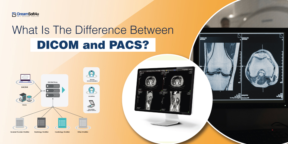
In the health sector, digitalization has resolved numerous problems. The prime example is easy access to patient information. Physicians could not access patient information like MRI scans or X-rays easily because they were performed at other hospitals. This is where DICOM (Digital Imaging and Communications in Medicine) Medical Imaging Software Development comes in as a single-window solution.
This software complies with an international standard that allows physicians to store, share and manage patient information with complete security and convenient accessibility, irrespective of device or location.
The worldwide medical imaging software market is anticipated to expand at a CAGR of 7.84% to $12.76 billion by 2030. It visually represents how rapidly its use is growing in the healthcare industry.
Now, all the clinics and hospitals are gearing up to create DICOM medical imaging software to be implemented within their existing system and handle massive amounts of patient data with tight compliance without any hassles. But it is not so easy!
Developing DICOM medical imaging software is a challenging task and has several factors to consider. No worries!
In this blog, we will understand everything about DICOM medical imaging software, its importance, key features and how you can build a DICOM medical imaging software with its estimated cost of development.
By the end of this blog, you will know exactly how you can build a DICOM medical imaging software and streamline workflows.
Table of Contents
ToggleWhat is DICOM Medical Imaging Software?
Medical imaging software DICOM Trends is a computer application. It assists physicians in interpreting medical images such as X-rays, MRIs, CT scans, ultrasounds, and PET scans. It helps physicians visualize precise information about diseases and suggest proper cures.
This application employs very advanced algorithms for the restoration of images, targeting sensitive areas, and helping doctors reach the correct conclusion. It is used extensively in radiology, cardiology, oncology, etc.
To enhance efficiency, this software integrates advanced technologies, including:
- AI for anomaly detection: Identifies potential health issues automatically.
- Machine Learning for image segmentation: Helps highlight specific areas of concern.
- Image enhancement techniques: Filters, contrast adjustments, and noise reduction improve clarity.
- 3D reconstruction: Creates volumetric models of organs and tissues.
- DICOM standard support: Ensures seamless image sharing between systems.
- Cloud technology: Enables remote access and data storage.
- VR & AR: Provides interactive, immersive visualizations for better analysis.
Through medical imaging software development, clinics and hospitals ease the burden of doctors, improve the accuracy of diagnosis, and minimize the chances of diagnostic mistakes.
Essential Features of Custom Medical Image Analysis Software Development
Some of the key features of medical imaging software are:
Image Quality Enhancement
High-resolution pictures are essential to make a proper diagnosis. This feature enhances medical image quality by removing noise, increasing contrast, and sharpening edges. It helps doctors clearly view even the slightest abnormalities. It makes the detection of diseases such as cancer, fractures, or infections easier. Better images enable faster, more accurate, and more reliable diagnosis.
Image Segmentation
Segmentation divides a medical image into distinct regions, such as organs, bones, or tissues. Segmentation allows physicians to concentrate on areas of concern, e.g., tumours or blood vessels, without distraction. AI-powered segmentation facilitates easy detection of abnormalities, measuring the extent of affected areas, and treatment planning such as surgery or radiation therapy. Segmentation is time-saving and accurate.
Image Registration
Medical imaging typically involves Physicians comparing scans taken at different points or from other machines (MRI, CT, X-ray, etc.). Image registration aligns these scans, so comparing the changes over time becomes more effortless. For example, a doctor can track how the size of a tumour grows or shrinks during treatment and hence make more informed decisions. This helps track disease progression or in the planning of treatments.
3D Reconstruction & 2D Visualization
2D pictures provide information, but 3D models offer a complete image of tissues and organs. This is useful for surgery, treatment planning, and seeing problems more clearly. For example, a 3D heart scan allows doctors to detect blockages better, as they can detect blockages more easily.
Quantification
Quantification translates pictures into numbers. It allows doctors to assess tumour size, measure blood flow, or analyze bone density. Numerical information helps track disease development, personalize treatments, and make precise diagnoses. Quantification is not always necessary; otherwise, image interpretation becomes subjective and may lead to mistakes.
AI-Driven Image Analysis
AI enhances medical imaging through the automatic detection of abnormalities such as lung nodules or brain tumours. AI can scan thousands of images in seconds and flag potential issues to be reviewed by radiologists. It not only saves time but also increases precision, reducing false negatives. AI helps identify patterns and detect diseases early, like Alzheimer’s or cancer.
Medical Data Management
Medical image management guarantees images are safely stored, easily accessible, and associated with patient records. It enables physicians to rapidly access previous scans, view previous and new photos side by side, and make decisions. Safe storage also monitors HIPAA and other data protection regulations.
Diagnostic & Treatment Tools
Doctors need more than images; they need measurement and planning software. This ability has a range of tools like tumour measurement, 3D surgical modelling, and radiation therapy planning. For example, a surgeon can use a virtual 3D model of the brain to plan an operation before walking into the operating room. Such tools reduce risks and improve patient outcomes.
EMRs/EHRs/PACS/RIS Integration
Smooth integration with Radiology Information Systems (RIS), Picture Archiving and Communication Systems (PACS), and Electronic Health Records (EHRs) is needed for an unobstructed workflow. It allows physicians to see images along with lab reports, medications, and medical history in a single place. It saves time, reduces errors, and improves patient care.
Reporting & Documentation
Physicians must create reports from scans to present to other medical professionals. This feature automates report creation, supports annotation, and organizes records well. It provides brief, standardized, and precise medical records to make follow-ups and treatment planning more manageable.
How to Build a DICOM Medical Imaging Software? 6 Simple Steps

The following are six simple steps to develop medical imaging software:
Step 1. Requirement Analysis
Take a step back before you go into development and figure out what your software actually has to do. Interview doctors, radiologists, and hospital staff.
Please find out about their everyday needs with medical images. Do they require improved storage of images? Improved analysis? Equipment driven by AI?
By knowing the requirements of your organization, you can develop more solution-focused software to deliver the required outcomes.
Additionally, ensure your software is compliant with required healthcare regulations such as HIPAA and GDPR. Gaining good knowledge at this point will save you money and time in the future.
Step 2. Choose the Right Tech Stack and Essential Features
Choose the right tools to develop your software. Choose programming languages and platforms that are secure and friendly to medical imaging. Think about the most critical features your users need like AI analysis, 3D imaging, or easy integration into hospital systems (EHRs, PACS). The right tech and features will make your software effective, reliable, and easy to use.
Step 3. Choose a Monetization Model
Before building your software, choose how it will make money. There are several options:
- Subscription Model: Charge users a monthly or yearly fee. It is ideal for hospitals and clinics needing regular access.
- Pay-Per-Use: Users pay per scan or image analysis. Great for smaller clinics or occasional users.
- Licensing Model: Sell the software for a one-time fee. It often includes support and updates.
- Commission-Based: Charge a fee per transaction. Works well for AI-assisted diagnostics or remote imaging services.
- Value-Based Pricing: Charge based on the benefits provided. Example: saving doctors time or improving diagnosis accuracy.
Pick the model that fits your users and business goals. A smart pricing strategy ensures long-term success.
Step 4. Hire an Experienced Software Development Partner
All companies do not have technical experts. That is when outsourcing to a well-experienced software development organization is a suitable option. They have the knowledge, expertise and experience to create tailor-made medical imaging software development for your business. Some important aspects you have to research thoroughly when choosing a software development company are experience, portfolio, technical know-how, and post-support and maintenance.
Step 5. Design, Develop and Test
It’s now time to hand the project over to the professional and make it a tangible thing. It begins with the designing, where the developers design an interface that’s easy to use for doctors and technicians.
Then, there is development, where engineers write the code, DICOM standards are incorporated, and efficient data processing is ensured. The software has to manage image uploading, analysis, and secure storage.
After construction, thorough testing has to be performed. The team conducts bug, security, and system performance testing. They also test the software to ensure it integrates smoothly with hospital systems such as EHRs and PACS.
Step 6. Launch and Maintain
After your software is ready, deploy it to your users. Train medical staff to use it. But do not leave it there – watch how it works, gather feedback, and push out updates to fix problems and add new functionality. Healthcare technology is constantly evolving, so continuing improvements will render your software functional and relevant.
How Medical Image Analysis Software Works?
Here’s how medical imaging software works:
Step 1: Image Capture
Medical devices like X-rays, MRIs, and CT scans take pictures of the body. These images help doctors find diseases or injuries. The software stores them in a digital format for further analysis.
Step 2: Image Processing
The software improves image quality. It removes noise, adjusts brightness, and enhances details. This makes it easier for doctors to spot issues.
Step 3: Image Analysis
AI and intelligent algorithms study the images. The software highlights problem areas and detects patterns. It helps doctors find diseases faster and more accurately.
Step 4: Visualization & Interpretation
Doctors view the images on a screen. They can zoom in, rotate, and see details in 2D or 3D. This helps them make better diagnoses and treatment plans.
Applications of Custom Medical Image Analysis Software
These are some of the applications of medical imaging software:
Diagnosis
Doctors use medical imaging to detect diseases like fractures, tumours and heart problems. X-rays, MRIs and CT scans enable them to see inside the body and detect abnormalities in seconds.
Treatment Planning
The software assists physicians in scheduling treatments such as surgery or radiation. It displays images of tissues and organs in tremendous detail, enabling accurate and individualized treatment plans.
Monitoring Disease Progression
Doctors track how a disease changes over time by comparing new and old images. They can then verify if a treatment is working and adjust accordingly.
Research and Education
Medical imaging is helpful for research and medical education. Students can learn human disease and anatomy through images, and researchers use images to create new treatments and diagnostics.
Early Disease Detection
Screening schemes use imaging to detect disease at an early stage. For example, mammograms identify breast cancer before symptoms are realized, resulting in earlier treatment and improved survival.
Personalized Medicine
Physicians interpret images to derive treatments tailored to specific patients. This provides more effective outcomes by taking into account individual factors such as tumour size, location, and human anatomy.
Who Needs Medical Image Analysis Software Development?
Let’s think about who needs medical imaging software:
- Hospitals: They use it to store, display, and handle medical images. It allows physicians to diagnose faster and improves the coordination of treatment. It also offers data privacy and compliance.
- Clinics: Clinics use it for rapid and accurate diagnosis. It simplifies image storage, reduces cost, and accelerates workflow. Physicians have immediate access to patient scans.
- Research Institutes: Scientists use it to analyze diseases and come up with solutions. AI applications help to decode viruses, genes, and the body’s functioning, leading to health advances.
- Imaging Centers: It enhances the accuracy of diagnostic tests. Imaging centres can handle enormous amounts of patient data securely at reduced costs and with maximum efficiency.
- Veterinary Clinics: The veterinarians use it to diagnose and treat animals. It speeds up imaging for illness and injury, improving animal care.
- Medical Device Manufacturers: They use it to enhance their medical devices. It helps in testing new technology, accuracy, and healthcare standards.
- Telemedicine Providers: It enables remote diagnosis. Patients can send scans to doctors from anywhere, which increases access to health care.
How Much Does It Cost to Build a DICOM Medical Imaging Software?
The average cost of building a DICOM medical imaging software ranges between $3,000-$300,000. However, the actual cost can vary based on several factors that affect price.
Here are some key factors that can affect the cost of medical imaging software development:
Complexity of Features
Advanced features like EHR integration or AI analysis increase costs—the more complex the features, the higher the price.
Customization Level
Essential tools are cheaper, while advanced customization, such as enhanced image viewing or analytics, requires more time and money.
Security Measures
DICOM software must comply with healthcare regulations. Stronger security (encryption, access controls) adds to development costs.
Scalability & Infrastructure
If the software must handle future growth, it needs robust servers, storage, and network capacity. Planning for scalability increases expenses.
Integration with Existing Systems
Connecting with hospital systems (EHR, HIS) adds complexity. Planning integration early helps control costs.
Multi-Standard Support
To work with different healthcare systems, the software may need to support multiple standards, making development more expensive.
Development Team & Location
Experienced developers cost more, especially in regions with high labour rates. Hiring skilled DICOM viewers specialists increases overall costs.
Challenges of Medical Imaging Software Development
Here are some significant challenges of medical imaging software development:
- Regulatory compliance: Health imaging software needs to be HIPAA, GDPR, and FDA law-compliant to offer protection for patient data. Strong encryption, two-factor authentication (2FA) and role-based access are emphasized. Consultation with regulatory experts avoids fines.
- System Integration: Hospitals use different IT systems like DICOM & PACS, EHRs and RIS. The software must support DICOM, HL7 and FHIR for smooth integration. Middleware helps bridge gaps.
- Performance & Scalability: Large CT and MRI images are processed slowly. Solutions include lossless compression, multithreading, and cloud storage.
- Big Data Management: Medical data storage requires powerful databases. Using backup systems and auto-recovery prevents data loss.
- Cybersecurity Risks: Healthcare software faces cyberattacks. Requires constant monitoring, security patches, and updates to protect patient data.
Future of Medical Imaging Software Development in Healthcare
Medical imaging is evolving faster, and more accurate ways to diagnose and treat patients are being found. Here is what is coming next:
1. AI-Powered Diagnostics
Artificial intelligence (AI) will help analyze images faster and detect diseases earlier with greater accuracy.
2. IoT for Real-Time Monitoring
Connected devices will allow instant image capturing and remote patient monitoring, improving diagnosis and treatment.
3. Cloud-Based Storage
Cloud technology will provide secure and scalable storage, making medical images easier to access from anywhere.
4. Stronger Cybersecurity
Advanced security features will protect patient data from cyber threats, ensuring privacy and compliance.
5. 3D and Virtual Reality Imaging
Doctors will use 3D imaging and VR tools to get detailed, interactive views of organs, helping with better diagnosis and surgery planning.
Planning for Medical Imaging Software Development?
We’ve a team of professionals who offers tailored solutions
Conclusion
Developing a DICOM medical imaging solution is a game-changer for healthcare organizations. It improves diagnosis, treatment, and patient data management. However, building such software from scratch is a complex thing, and there are several things to consider. We hope this guide helps you understand the role of medical imaging software development in healthcare and how you can build medical imaging software with an estimated cost. Now, it’s your turn to hire an experienced software development firm and let the professionals build your medical imaging software within budget and on a timeline.
DreamSoft4U is a trusted software development firm which has been offering customer medical imaging software development for over 20 years. We have a team of skilled experts who provide tailored solutions for healthcare businesses of all sizes at an affordable price. Contact us today and turn your vision into reality!
Medical Imaging Software Development – FAQs
Q1. Why is medical imaging software essential?
It helps doctors see and analyze medical images. Such as X-rays and MRIs more clearly. It improves accuracy, speeds up diagnosis and reduces workload. Plus, it keeps patient data safe and organized.
Q2. What are the key features of DICOM medical imaging software development?
- Better Image Quality: Clears up details for accurate diagnosis
- AI Analysis: Spots issues like tumours automatically.
- 3D Imaging: Gives a full view of organs and tissues.
- Data Management: Stores and organizes images securely.
- System Integration: Works with hospital records (EHRs, PACS, RIS).
- Easy Reporting: Automates medical reports.
Q3. Who needs DICOM medical imaging software?
- Hospitals & Clinics
- Imaging Centers
- Research Labs
- Veterinary Clinics
- Telemedicine Providers
Q4. How does AI improve medical imaging?
AI helps doctors by quickly spotting issues in scans. It highlights tumours, improves image quality, and detects diseases early. It also saves time by analyzing images in seconds.
Q5. Can medical imaging software integrate with hospital systems?
Yes! It integrates with EHRs, PACS and RIS. So, doctors can view images alongside medical records. This enhances patient care in terms of efficiency and speed.
Q6. How long does it take to build medical imaging software?
It depends on the features. A basic version takes a few months, while an advanced AI-powered system can take a year or more.


